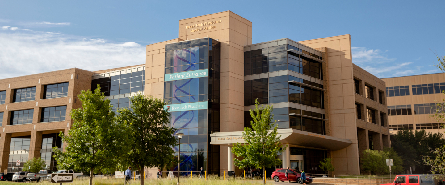
Abilene Core Laboratory Equipment
The Jerry H. Hodge School of Pharmacy on the Abilene campus offers the following core facilities and equipment: Aperio Scanscope, BD LSR Fortessa Cell Analyzer, Flow Cytometer Cell Sorter BD FACSAria, Nikon Microscope A1, IVIS Spectrum In Vivo Imaging System and an RS2000 Irradiator.
Immunotherapeutics and Biotechnology
-
Create high-quality digital slides with the ultra-compact Aperio CS2 image capture device – from your desktop. With a five-slide capacity and 20X and 40x magnification capabilities, the Aperio CS2 is a highly reliable workhorse for medium-volume sites.
Image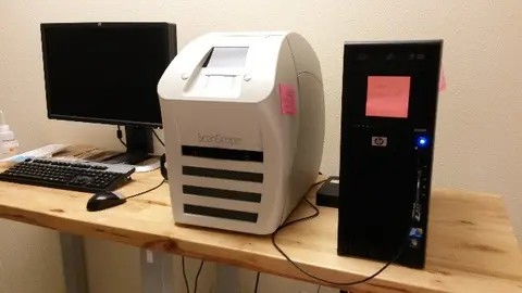
Aperio CS2 is the ideal tool for preparing slides and data to be shared with other team members for collaboration. Remote assessment of digital slides can improve study turnaround time and ease of access for team members.
A compact, small footprint, high image quality whole slide line scanning instrument, Aperio CS2 occupies only 1.6 square feet of bench top space.
Key Features:
- External sharing and collaboration (institution to institution)
- Internal sharing (Hub & Spoke Network)and collaboration
- eIHC image analysis
- Contract research
- Tissue microarrays
View Aperio Image Analysis User’s Guide
Location:
Texas Tech University Health Sciences Center
School of Pharmacy - 2512
1718 Pine St.
Abilene, TX 769601Contact Information:
Dr. Maciej Markiewski
Office: Abilene Campus Room 2513
Phone: 325-696-0430
Email: maciej.markiewski@ttuhsc.eduBhaumik Patel
Office: Abilene Campus Room 2503
Phone: (325) 696-0484
Email: bhaumik.patel@ttuhsc.edu -
The BD LSRFortessa™ cell analyzer offers the ultimate in choice for flow cytometry, providing power, performance, and consistency. Designed to be affordable and expandable, the BD LSRFortessa has the flexibility to support the expanding needs of multicolor flow cytometry assays. The BD LSRFortessa analyzer can be configured with up to 4 lasers—blue, red, violet, and UV—which enables the detection of up to 18 colors simultaneously. The BD LSRFortessa may be upgraded subsequently with additional or new lasers from BD, as future requirements dictate.
Image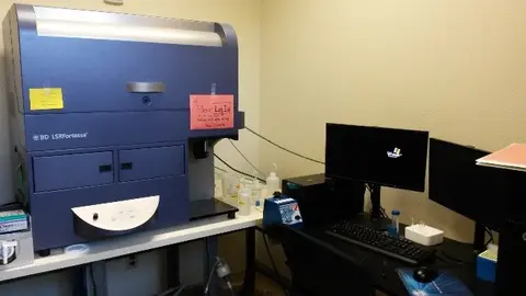
Lasers:
- 405 Violet Laser
- 488 Blue Laser
- 640 Red Laser
Key Features:
The fluidics system is pressure driven. Hydrodynamic focusing forces sample cells through the cuvette flow cell, where they are interrogated. The flow cell is in fixed alignment with the laser and gel-coupled to the collection optics. This design ensures that the laser is precisely focused on the sample stream and the maximum amount of emitted light can be collected for added sensitivity in multicolor applications. Fixed alignment also minimizes startup time, improves experiment-to-experiment reproducibility, and enables automated daily quality control.
The optics system consists of laser excitation optics that illuminate cells in the sample, and collection optics that direct light scatter and fluorescence signals through spectral filters to detectors. Innovative designs for both the excitation optics and collection optics in BD LSRFortessa systems reduce excitation losses and optimize collection efficiency for increased sensitivity and resolution.
View BD LSRFortessa Cell Analyzer User’s Guide
Location:
Texas Tech University Health Sciences Center
School of Pharmacy - 2414
1718 Pine St.
Abilene, TX 769601Contact Information:
Dr. Laurence Wood
Office: Abilene Campus Room 2514
Phone: 325-696-0431
Email: laurence.wood@ttuhsc.eduBhaumik Patel
Office: Abilene Campus Room 2503
Phone: (325) 696-0484
Email: bhaumik.patel@ttuhsc.edu -
The BD FACSAria cell sorter sets a new standard for high-performance flow cytometry. Based on an entirely new design in instrumentation, the BD FACSAria instrument is the first benchtop sorter that incorporates a fixed-alignment cuvette flow cell. The numerous technological advances embodied in the BD FACSAria cell sorter reduce the cost of owning a highspeed sorter and accelerate research by providing unparalleled, easy-to-use, high-performance sorting and analysis.
With 3 lasers, it can detect up to 11 fluorochromes, for a total of 13 parameters:
- 488 nm: 7 detectors (SSC + 6 fluorochromes)
- 633 nm: 3 detectors
- 405 nm (violet): 2 detectors
Key Features:
- High-speed cell sorting
- 3 air-cooled lasers (blue: 488, red: 633, and violet: 407 nm)
- Digital acquisition rates of up to 70,000 events/second
- Multicolor analysis (up to 9 colors)
- Two- and four-way bulk sorting into tubes and 96-well plates
- Optical filters to detect fluorophores in the following nine channels:
- Digital Fluorescence-activated cell sorter
View Flow Cytometer Cell Sorter BD FACSAria User Manual
Location:
Texas Tech University Health Sciences Center
School of Pharmacy - 2408
1718 Pine St.
Abilene, TX 769601Contact information:
Dr. Laurence Wood
Office: Abilene Campus Room 2514
Phone: 325-696-0431
Email: laurence.wood@ttuhsc.eduBhaumik Patel
Office: Abilene Campus Room 2503
Phone: (325) 696-0484
Email: bhaumik.patel@ttuhsc.edu
-
The A1 is Nikon's powerful fully-automated confocal imaging system, capable of capturing high-quality confocal images of cells and molecular events at high speed and enhanced sensitivity. Ideal for facilities with a broad range of users, the A1 has been designed with groundbreaking new optical and electronic technology innovations to provide unprecedented system quality and flexibility.
- Equipped with 4 lasers 405, 488, 561, and 635nm.
- High precision motorized-focus, and image capture in 3D and many more features.
Image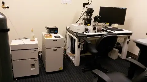
Key Features:
- A high-speed resonant scanner allows imaging of intracellular dynamics at 30 frames per second (Fps). Moreover, image acquisition of 420 fps is also possible.
Simultaneous imaging and photo activation with the proprietary hybrid scanner reveal intermolecular interaction. Analysis software for FRAP and FRET is provided standard. - Fast spectral image acquisition for 32 channels at a maximum of 24 fps (512 x 32 pixels) is possible. New real-time spectral unmixing and the V-filtering function expand the range of use of spectral images.
Fluorescence efficiency is increased by 30 percent, and S/N ratio of images is also increased. With diverse new technologies such as the VAAS pinhole unit, superior image quality has been achieved.
View Nikon Microscope A1 Manual
Location:
Texas Tech University Health Sciences Center
School of Pharmacy - 2502
1718 Pine St.
Abilene, TX 769601Contact information:
Dr. Magdalena Karbowniczek
Office: Abilene Campus Room 2517
Phone: 325-696-0430
Email: magdalena.karbowniczek@ttuhsc.eduBhaumik Patel
Office: Abilene Campus Room 2503
Phone: (325) 696-0484
Email: bhaumik.patel@ttuhsc.edu -
IVIS from Perkin Elmer offers single-view 3D tomography for both fluorescent and bioluminescent reporters and has capability to illuminate either trans-illumination (from the bottom) or epi-illumination (from the top) fluorescent sources that can be analyzed in an anatomical context.
3D diffuse fluorescence tomography can be performed to determine source localization and concentration using the combination of structured light and trans illumination fluorescent images.
Key Features
· Twenty eight high efficiency filters spanning 430 – 850 nm makes it ideal for high-sensitivity in vivo imaging of fluorescence and bioluminescence
· High throughput (5 mice) with 23 cm field of view
· High resolution (to 20 microns) with 3.9 cm field of view
· Supports spectral unmixing applications leads to significant decrease in background signal
View IVIS Spectrum In Vivo Imaging System Manual
Location:
Texas Tech University Health Sciences Center
Abilene Campus – Room# 102P
842 Pine St.
Abilene, TX 769601Contact information:
Bhaumik Patel
Office: Abilene Campus Room 2503
Phone: (325) 696-0484
Email: bhaumik.patel@ttuhsc.edu -
RS 2000 specializes in providing non-isotope, ionizing irradiation. Ionizing radiation that is similar to the radioactive sources, cobalt-60 and cesium-137. RS 2000 utilizes Rad Source’s X-ray tube technology and RAD+ reflector to generate an efficient and uniform X-ray ionizing radiation field, to provide excellent irradiation effect, and good repeatability.
Image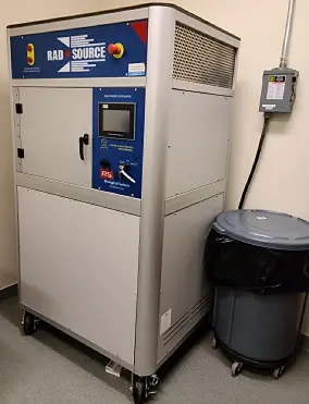
Key Features
- RBE equal to cesium and cobalt irradiators–no isotopes
- Irradiation in a filtered environment (no cross-contamination issues).
- Programmable techniques using touch panel screen.
- Complete dose mapping.
- PLC control system
View Irradiator: RS2000 Manual
Location:
Texas Tech University Health Sciences Center
Abilene Campus – Room# 100
842 Pine St.
Abilene, TX 769601Contact Information:
Bhaumik Patel
Office: Abilene Campus Room 2503
Phone: (325) 696-0484
Email: bhaumik.patel@ttuhsc.edu
Amarillo Core Laboratory Equipment
The Mass Spectrometry Core Lab is committed to advancing scientific research and promoting a safer and healthier world through cutting-edge analytical services and expert advisory support. Our mission is to foster innovation, deliver timely results, and maintain the highest standards of quality using state-of-the-art instrumentation and scientific excellence. By collaborating with researchers across diverse fields, we aim to drive discoveries, empower decision-making, and contribute to the global pursuit of knowledge.
The mission of the Amarillo Imaging Core is to provide researchers across West Texas and beyond access to cutting edge instruments and imaging service, covering the range from small animal live imaging to optical super resolution microscopy.
Amarillo Mass Spectrometry Core Instruments
Contact us to request more information:
Ulrich Bickel, M.D.
Associate Dean of Sciences
ulrich.bickel@ttuhsc.edu
(806) 414-9236
Dhaval Patel, M.S., MBA
Research Lab Manager
Dhavalkumar.patel@ttuhsc.edu
(806) 414-9094
-
Image
About:
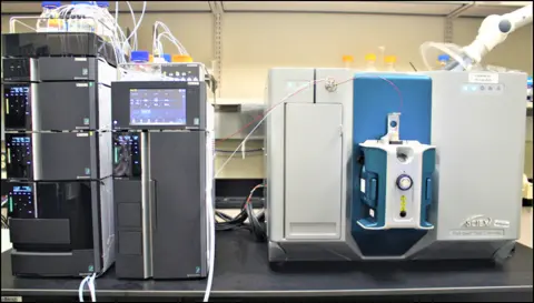
This instrument offers a superior quantitative result with its Triple Quadrupole (QQQ) technology as well as optional Linear Ion Trap (LIT) functionality. This instrument mostly used in our core facility for the Multiple Reaction Monitoring (MRM) of small molecules quantitative analysis which provides highly sensitive and reproducible results from complex matrices such as plasma, urine, tissues, etc. The QTRAP technology offers additional scan type such as MRM3 (MS/MS/MS), Enhanced Product Ion Scans (EPI) and many more that offers additional benefits of septicity and semi-qualitative analysis through its unique QTRAP technology.
Whether your research emphasizes on drug discovery and development, Biomarker study, food and beverages analysis or searching an answer from complex analysis, the 5500 & 7500 QTRAP system is equipped to handle those challenges.
Key Features:
- D Jet ion guide: Capture more of the ESI plume and retain more important ions. The D Jet ion guide concentrates samples and removes gas molecules and neutral ions.
- QTRAP system: Gain additional QTRAP functionality. Pairing conventional MRM workflows and Enhanced product ion scans enable improved confidence in acquired data. MRM3 workflows push quantification levels through matrix interferences.
- Detection: Attain lower levels of quantification. The precise and robust engineering of the ion rail allows consistent and reproducible analysis time after time, by focusing the crucial ions you need for your workflow.
- Ionization source: Execute fast interchanges between high flow and low flow to adapt to your workflow needs. The OptiFlow Pro ion source introduces a new modularity feature and incorporates the reliability and efficiency of the legendary Turbo V ion source.
- E Lens probe: Get a stronger gradient around the orifice and protect your precious sample. With E Lens Technology, the SCIEX Turbo V Ion Source geometry is enhanced in the new OptiFlow Pro Source, which focuses transmission of the ESI plume into the orifice of the system.
Location: Texas Tech University Health Sciences Center, Amarillo Research Building, 1406 Coulter St. Room 2303
Amarillo, TX 79106 -
Image
About:
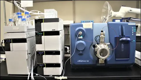
The UHPLC/MS/MS system is composed of a highly sensitive AB SCIEX QTRAP 5500 mass spectrometer interfaced to a Shimadzu NEXERA Ultra High Performance Liquid Chromatograph (UHPLC). The NEXERA UHPLC offers pressure range up to 19000 psi and is suitable for both conventional and fast analysis in combining use of small particle column. The QTRAP 5500 is a hybrid Triple Quadrupole Linear Ion Trap mass spectrometer, offering some very unique features.
- Mass/charge range of 5-1250 Da in triple quadrupole mode and 5-1000 Da in linear ion trap mode.
- Operating software : Analyst 1.6.3; Quantitation software: MultiQuant 3.0.3.
- Multiple reaction monitoring (MRM) quantitation using triple quadrupole system.
- Both qualitative and quantitative analysis in single run.
- High sensitivity and relatively high resolution full-scan MS, MS/MS, and MS3 in Linear Ion Trap mode.
- Triple quadrupole precursor ion and neutral loss scans.
- Metabolite identification facilitated by LightSight 2.2.1and ACD/labs MS processor.
Location: Texas Tech University Health Sciences Center, Amarillo Research Building, 1406 Coulter Street, Room 2303, Amarillo, TX 79106
-
Image
About:
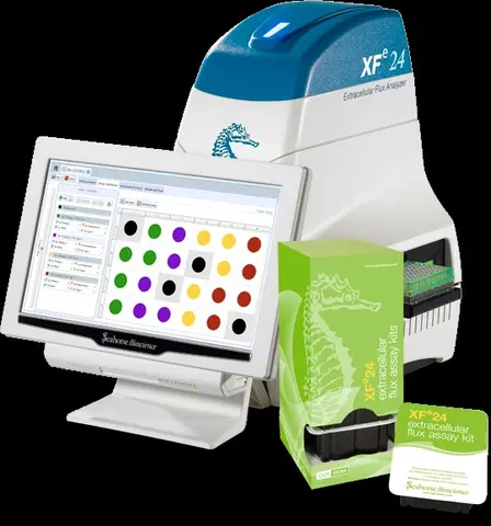
Monitors bioenergetics of live cells, in real time, in a microplate.
The two major pathways to produce energy, mitochondrial respiration and glycolysis, involve cellular consumption of oxygen and efflux of protons, respectively. Seahorse XF technology uses label-free sensors to detect extracellular changes in these analytes in order to measure rates of cellular respiration, glycolysis, and ATP production. Cells are seeded in the assay wells of the custom 24-well XF microplate at a confluency of 50-90%. Suspension cells are attached to the well bottom to maximize sensitivity.Forms a microchamber and calculates rates of extracellular flux in minutes.
The instrument lowers the cartridge probes into the assay wells. The sensors are positioned 200 microns above the well bottoms, forming transient microchambers of approximately 7 microliters (28 for islet plates). As the oxygen and pH levels change, the changes in the sensors are read by the instrument. Measurements are typically made for 3 minutes and rates are calculated automatically. Upon completion of this measurement period the probes are raised, allowing the extracellular medium to come back to baseline conditions.Injects up to 4 compounds to test responses or interrogate biology in real time.
The sensor cartridge also contains ports (4 per well) to enable injection of modulators into the cell wells during the assay. When specified by the instrument protocol, the system injects compound "A" into the assay wells and performs a gentle mixing step to ensure distribution of the compound throughout the assay medium, followed by a wait period. All wells are processed in this manner simultaneously. Subsequent measurement cycles, any additional injections specified by the protocol, and rate calculations are performed automatically.Key Features:
- Live cell, real-time analysis of cellular energy metabolism platform in 24-well format
- Multiple parameters reported from each assay well, including OCR, PER or ECAR, and ATP product rates
- Large well size and transient microchamber to provide the capacity for larger and/or more metabolically active samples
- Robust response with as few as 10,000 cells per well in the custom 24-well plate
- Compatible with 3D study models such as islets and small organism (for example, zebrafish)
- Four-port injection system with automated mixing feature for assessing immediate cellular responses to substrates, inhibitors, and other compounds in real time
- Wide operation environment temperature of 4 to 30 °C, allowing the analyzer to maintain internal assay temperature of 16 to 42 °C for compatibility with a variety of samples
- Data files compatible with the web-based analytics tool, Seahorse Analytics
Location: Texas Tech University Health Sciences Center, Amarillo Research Building, 1406 Coulter Street, Room 1207A, Amarillo, TX 79106
-
Image
About:
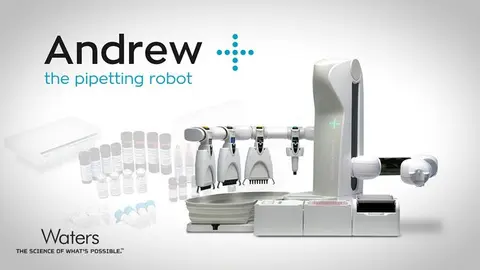
The Andrew+ is a state-of-the-art pipetting robot designed to revolutionize laboratory workflows. With its fully automated pipetting capabilities, it offers unparalleled precision and accuracy in liquid handling tasks. Equipped with a wide range of Domino Accessories and Andrew Alliance electronic pipettes, it enables not only routine pipetting but also complex manipulations with ease.
By seamlessly executing OneLab protocols, the Andrew+ eliminates the need for laborious manual procedures, reducing human error and increasing efficiency. This cutting-edge instrument empowers researchers to swiftly transition to error-free, robotic workflows, saving valuable time and resources.
In addition to its exceptional pipetting performance, the Andrew+ liquid handler boasts an impressive array of capabilities. It can seamlessly transport tubes and plates, streamlining sample preparation and processing. Furthermore, with its vacuum-assisted functionality, it can perform advanced techniques like solid-phase extraction, expanding its utility in diverse experimental workflows.
The Andrew+ represents a significant advancement in laboratory automation, empowering scientists to focus on higher-level tasks while achieving consistent and reproducible results.
How does it look: Please watch the following video.
https://ttuhsc.box.com/s/k1z95en5yvci0t8g7npj4ul60aph2ba7
Want to learn more about it? Please refer https://www.andrewalliance.com/pipetting-robot/#
Amarillo CPRIT Imaging Core Instruments
The mission of the Amarillo Imaging Core is to provide researchers across West Texas and beyond access to cutting edge instruments and imaging service, covering the range from small animal live imaging to optical super resolution microscopy.
Note About Citations: Please acknowledge the CPRIT Imaging Core in all publications or grant proposals as follows: "...funded by the Core Facility Support Award (grant number RP200572) from the Cancer Prevention and Research Institute of Texas (CPRIT) to the Imaging Core, Texas Tech University Health Sciences Center at Amarillo."
Contact us to learn more:
Ulrich Bickel, M.D.
Associate Dean of Sciences
ulrich.bickel@ttuhsc.edu
(806) 414-9236
Zijuan "Amy" Liu, Ph.D.
Research Lab Manager
zijuan.liu@ttuhsc.edu
(806) 414-9199
Alex Mdzinarishvili, Ph.D.
Research Lab Manager
Alexander.Mdzinarishvili@ttuhsc.edu
(806) 414-9196
-
-
Image
About:
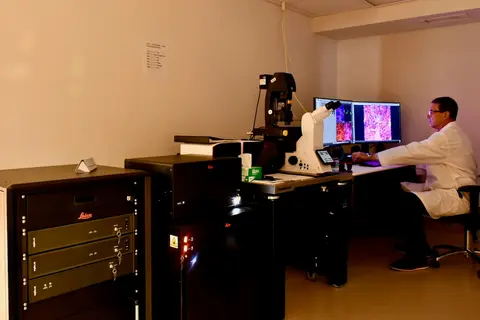
Spectral confocal microscope with optical super resolution capability (Stimulated Emission Depletion, STED) enabling resolution down to ~30 nm. FAst Lifetime CONtrast (FALCON) maps fluorescence lifetime, enhancing contrast in combination with STED (tau-STED). The system is further equipped with an optional on-stage live cell imaging chamber (Okolab) and an AI-based quantitative image analysis package (AIVIA).
Key Features:- DMi8 inverted microscope stand
- Immersion lenses for super resolution imaging (< 50 nm)86x (water), 93x (glycerol), 100x (oil)
- White light laser, 440nm – 790 nm, 405 nm UV laser
- Stimulated Emission Depletion (STED) module for optical super resolution
- STED lasers 592 nm, 660 nm, 775 nm
- 4 hybrid detectors, resonant scanner, transmitted light detector
- Fluorescence lifetime imaging (FALCON), tau STED.
- On-stage live cell incubation system (Okolab) for normoxia, hypoxia, or hyperoxia
- LAS X Control and Analysis Software
- Deconvolution (Lightning Expert)
- AIVIA 3D suite
-
-
About:Unique small animal imager that it operates at near infra-red (INR) to infrared (IR) wavelengths and avoids the various complications associated with absorption scattering and the autofluorescence of light in living tissues. Using the first (NIR) and second biological window (NIR-II) from 900-1620 nm, the IR VIVO provides multispectral or hyperspectral infrared imaging capabilities for in vivo studies on small animals including rats.
Image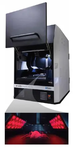
-
About:
The Livecyte Kinetic Cytometer is a complete imaging and analysis system optimized for label-free long term, non-invasive monitoring of live cells. It utilizes Ptychographic Quantitative Phase Imaging (QPI), fluorescence and bright field imaging. Ptychographic QPI allows robust automatic tracking and behavioral analysis of a significant number of individual cells within heterogeneous cell populations without labels. Unique morphological, temporal, and dynamic phenotypic data make it simple to gain behavioral information of individual cells of cell clusters through live cell imaging. Achieve more efficient imaging by using every well in any plate, up to 96 well format.
Image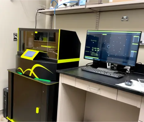
-
About:
In live cells and tissue imaging, a key technology is two photon laser scanning microscopy (TPLSM). It minimizes phototoxic effects and offers the possibility to image deep into tissues.
Key Features:
- Simultaneous excitation imaging with two-wavelength IR laser
- Deep in vivo imaging with super high-sensitive GaAsP NDD
- Ultrahigh-speed imaging of up to 420 frames per second
- Multi-color in vivo imaging
- Deep specimen imaging with non-descanned detectors (NDD)
- Nikon's high-NA objectives are ideal for multiphoton imaging
- Auto laser alignment when changing multiphoton excitation wavelength
- Two types of scanning head enable high-speed, high-quality imaging
-
NIS-Elements C acquisition and analysis software
Image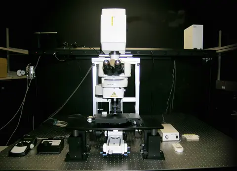
Dallas Core Laboratory Equipment
Contact us to learn more:
Ron Hall, Pharm.D.
Associate Professor
Ronald.Hall@ttuhsc.edu
Carlos Alvarez, Pharm.D.
Associate Professor
Carlos.Alvarez@ttuhsc.edu
-
A Sciex QTRAP® 5500 which is interfaced with a UHPLC, nano-LC or micro-LC system.
-
A Sciex QTRAP® 6500+ which is interfaced with a UHPLC, nano-LC or micro-LC system.
-
A Sciex TripleTOF™ 5600 which is interfaced with interfaced with a UHPLC, nano-LC or micro-LC system.
-
The Center’s UHPLC-MS/MS systems can be interfaced with Sciex’s SelexION™ which provides the unique capability to separate analytes with identical molecular weights and retention times.
Lubbock Core Laboratory Equipment
Lubbock is home to multiple core facilities and equipment administered by the TTUHSC, the TTUHSC School of Medicine Cancer Center, and TTU.
Texas Tech University Health Science Center
Image Analysis Core
The Imaging Analysis Core Facility provides state-of-the-art instrumentation for high-resolution light microscopic imaging. Expert advice is available for the design of experiments involving these microscopy techniques; and for the development of grants and manuscripts involving high-resolution light microscopic technologies.
Molecular Biology Core
The Molecular Biology Core Facility supports experimental needs in molecular biology, ranging from quantitative PCR, routine bacterial cell growth, monitoring of mammalian cell growth, advanced approaches for cell transfection, monitoring protein and gene expression, flow cytometry and cell sorting capabilities.
TTUHSC School of Medicine Cancer Center
The School of Medicine Cancer Center offers multiple core laboratories including the West Texas Pharmacology Core.
The West Texas Pharmacology core facility provides the most up-to-date technologies and expertise in clinical pharmacology in drug development and clinical trials. The core is funded by CPRIT to enable preclinical and clinical pharmacology studies in pediatric cancer drug development and offers the use of a Sciex Triple Quad 6500, Waters Arc HPLC systems (with DAD and FLD), and Shimadzu L20 HPLC.
Full List of Cancer Center Core Laboratories >
Texas Tech University
Center for Biotechnology and Genomics
The center provides instrumentation, support and training in various aspects of modern biotechnology including spectrophotometry, protein identification, protein purification molecular interaction, DNA applications and functional genomics.
College of Arts and Sciences Microscopy (CASM)
CASM supports microscopy and cell cytometry research through the use of techniques of transmission electron, scanning electron, laser confocal or epifluorescence microscopy. Trained users are given independent access to the instruments they require.
Texas Tech Neuroimaging Institute (TTNI)
TTNI is a multi-user neuroimaging facility that promotes cutting-edge interdisciplinary research. It provides researchers with brain and body imaging technologies including structural (MRI), functional magnetic resonance imaging (fMRI), magnetic resonance spectroscopy and diffusion tensor imaging and techniques, including multimodal data fusion of EEG, fMRI, and DTI data.
Lubbock Imaging Analysis Core Facility
The Imaging Analysis Core Facility provides state-of-the-art instrumentation for high-resolution light microscopic imaging. Expert advice is available for the design of experiments involving these microscopy techniques; and for the development of grants and manuscripts involving high-resolution light microscopic technologies.
To use this equipment, contact Dr. Grozdanov at imagecore@ttuhsc.edu or phone: +1(806)743-4624
-
Image
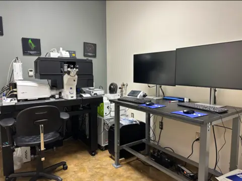
The Zeiss LSM 980 system is a highly advanced confocal microscope designed to enhance both sensitivity and ease of use. Its innovative filterless internal detector layout enables versatile multichannel imaging, complemented by a four-channel GaAsP array that delivers exceptional signal-to-noise ratios for quantitative analysis. The Airyscan 2 detector facilitates rapid and gentle super-resolution imaging based on pixel reassignment. It also facilitates flexible Multiplex modes that allow for the capture of larger fields of view and dynamic biological processes. Key features include: a confocal scanhead with two liquid-cooled MA-PMT detectors and four high-sensitivity GaAsP channels, full emission tunability, and integrated spectral acquisition capabilities. The Zeiss LSM 980 is equipped with solid-state diode laser lines ranging from 405 to 730 nm, including dual-channel near-infrared detectors sensitive to 900 nm. Additional components include an Observer 7 inverted microscope and motorized stand with an automated focus unit, an A.I. Sample Finder, long-lifetime fluorescence LED illumination, and incubation accessories for controlled environments. The system also includes Plan Apochromat objectives, ZEN 3.10 software for advanced imaging capabilities, a PC workstation, and an anti-vibration table, along with a ZEN 3.10 Desk Analysis Software for comprehensive and high-throughput image analysis.
-
Image
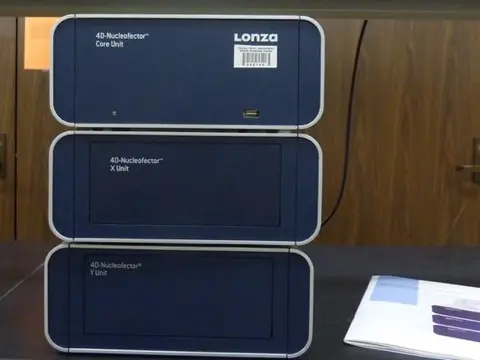
The 4D-Nucleofector Core X/Y (Lonza) system consists of two units: the X Unit, which is designed for transfecting cells in suspension and can handle various cell numbers and formats, and the Y Unit, which is used for transfecting adherent mammalian cells in 24-well plates. Both units are capable of transfecting cells with either DNA or RNA. For more information about the 4D-Nucleofector, please visit the Lonza website.
-
Image
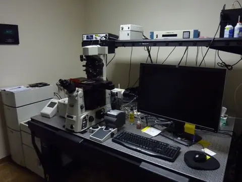
The Nikon T1-E microscope is a versatile imaging system featuring an A1 confocal module and STORM super-resolution capabilities. It can image both live and fixed cells and tissues using conventional or confocal microscopy techniques. Additionally, the STORM module provides super-resolution microscopy, which allows for extremely precise localization of fluorophores beyond the diffraction limit of light, achieving resolution up to ten times greater than standard optical microscopy. The system also supports spectral imaging and advanced fluorescent optical techniques, including TIRF, FRET, and FRAP. It is equipped with a high-resolution EMCCD camera and a stage-top environmental chamber for extended observation of living cells.
-
Image
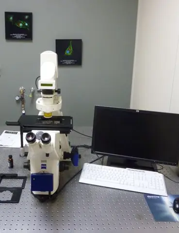
The Zeiss Axiovert 200 M microscope is a popular choice in the Image Analysis Core Facility. This inverted, fully motorized microscope features 10x, 20x, and 63x objectives for wide-field fluorescence microscopy. It has both color and monochrome Axiocam cameras attached, enabling the capture of various samples: color images (e.g., H&E-stained or peroxidase-stained) with the color camera, and low-intensity fluorescent images (e.g., YFP, GFP, RFP, and DAPI) with the monochrome camera. The microscope also supports phase contrast and differential interference contrast (DIC) imaging. Its stage includes a versatile holder that fits different sizes of cell culture dishes and plates, making it easy to quickly assess transfection efficiency.
-
Image
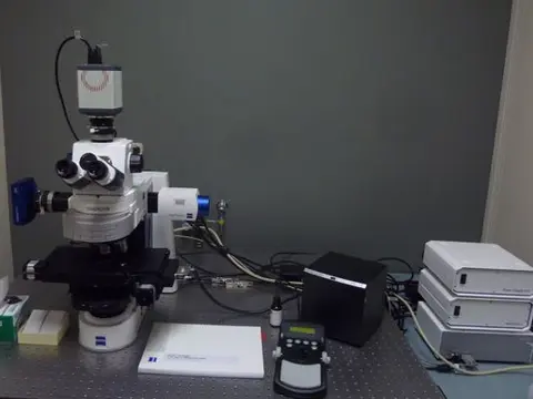
The Zeiss Axio Imager M2 microscope is a fully motorized system designed for brightfield and multi-channel fluorescent imaging. It features motorized x, y, and z stages and is equipped with the Zeiss Apotome system, which uses structured illumination to create sharp optical sections through thick specimens. Images are captured with a low-noise CMOS digital camera (ORCA-Flash4.0, Hamamatsu) with a resolution of 2048 x 2048 pixels. Additionally, the microscope is linked to Zeiss ZEN 3.5 Pro (Blue Edition) software, which offers advanced automated imaging and analysis tools to enhance your research.
-
Image
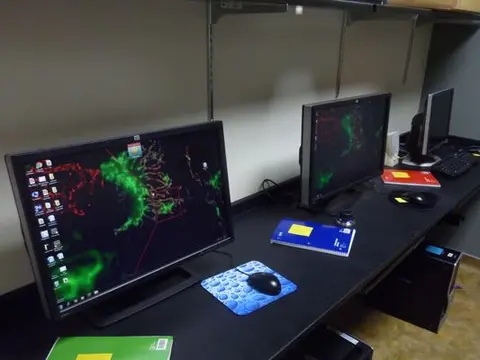
Our workstation area for data analysis is equipped with several computers dedicated to off-line analysis of digital images generated in the facility. Each computer is loaded with essential image analysis software, including Imaris. Imaris offers powerful tools for 3D and 4D image visualization and analysis, allowing you to track and quantify structures within your images. It supports advanced features such as automated object detection, spatial statistics, and detailed visualization of complex datasets. Whether you need to analyze cellular structures, track dynamic processes, or create detailed renderings, Imaris provides a comprehensive suite of tools to enhance your research.
Lubbock Molecular Biology Core Facility
The Molecular Biology Core Facility supports experimental needs in molecular biology, ranging from quantitative PCR, routine bacterial cell growth, monitoring of mammalian cell growth, advanced approaches for cell transfection, monitoring protein and gene expression, flow cytometry and cell sorting capabilities.
To use this equipment, contact Dr. Grozdanov at molcore@ttuhsc.edu or phone: +1 (806) 743-4624
-
Image
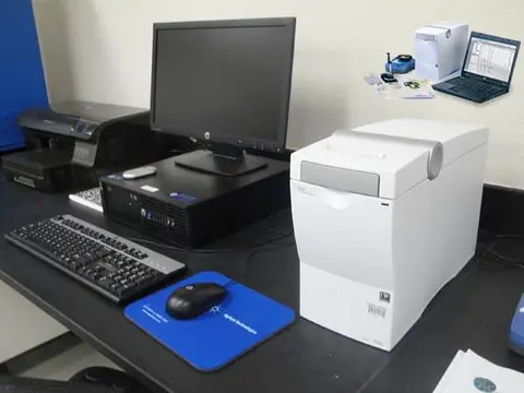
The 2100 Bioanalyzer uses microfluidics technology to analyze RNA, DNA, and protein samples. It's particularly useful for assessing RNA quality (RNA Integrity Number) before running microarrays, RNA-seq, or real-time quantitative PCR. The instrument analyzes each sample, creating a pseudogel image where nucleic acid or protein bands are sized and quantified. It requires only a minimal sample volume (4 µl) and provides fast, automated, and high-quality digital data. This replaces the need for time-consuming agarose gel or SDS-PAGE techniques. Sample preparation takes about 5 minutes, and digital results are available within 30 minutes. For more information, please visit the company's website.
-
Image
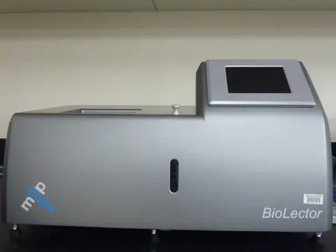
The BioLector (M2P Labs) is a versatile instrument designed for growing bacteria, yeast, or mammalian cells in suspension using a 48-well plate. It continuously monitors several key factors, including cell growth (biomass), fluorescence, acid production (pH), and oxygen consumption. The system also fully controls and adjusts CO2, oxygen, humidity, and temperature, making it ideal for rapidly optimizing growth conditions and conducting cytotoxicity assays. Additionally, the Multitron II shaker (used offline) complements the BioLector by enabling parallel experiments, thus allowing you to maximize efficiency and free up time on the BioLector. For more information, please visit the company's website.
-
Image
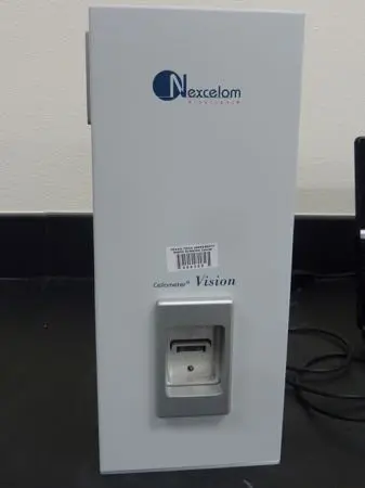
The Cellometer Vision Image Cytometry (Nexcelom Bioscience) system is designed for straightforward image cytometry with just a 20 µl sample. It can perform a variety of analyses, including cell cycle analysis, apoptosis, autophagy, cell proliferation, mitochondrial membrane potential, viability assays, morphology analysis, and cell counting. The instrument can monitor fluorescence from a range of dyes and fluorochromes, including GFP, FITC, Acridine Orange (for DNA and RNA), RFP, PI, and PE, across green and red spectra. It captures high-quality images using an integrated digital camera and a 20x objective lens. For more details about the instrument, please visit the company's website.
-
Image
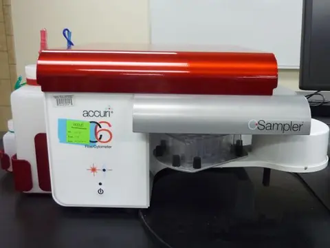
The BD Accuri™ C6 Plus Personal Flow Cytometer is a compact and lightweight flow cytometer, measuring 11 x 14.75 x 16.5 inches and weighing 30 lbs. It supports diverse applications, including immunology, cell and cancer biology, plant and microbiology, and industrial uses. Despite its small size, it offers powerful analysis capabilities with a blue and red laser and four fluorescence detectors optimized for common fluorophores like FITC, PE, PerCP, and APC. It accommodates 12 x 75 mm sample tubes and microcentrifuge tubes and can automate data collection from 48- and 96-well plates. The C6 Plus is ideal for studying various cellular functions, such as apoptosis, cell cycle, cell proliferation, mitochondrial membrane potential, immunoassays, cell phenotyping, cytosolic calcium and pH, and nitric oxide. For more information, visit the company's website.
-
Image
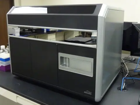
The ImageStream Mark II combines the rapid, statistical power of flow cytometry with the detailed insights of high-resolution microscopy. It offers high-resolution imaging in flow with 20x, 40x, and 60x objectives, four lasers (405 nm, 488 nm, 561 nm, 642 nm, 785 nm), and 12 imaging channels. Dual CCD cameras serve as detectors, allowing evaluation of various fluorochromes in non-adherent cells, whether alive or fixed. For more information, visit the company's website.
-
Image
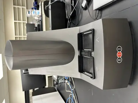
The MESO QuickPlex® SQ 120MM is a versatile and compact system known for its reliability and ease of use. It performs multiple tests on a single sample quickly, increasing productivity and saving sample volume. This instrument supports 96- and 384-well formats and can detect biomarkers in single or multiplex formats. It uses specialized kits for profiling various biomarkers and diseases, including cancer, cardiovascular issues, and neurodegenerative disorders. Equipped with an ultra-low noise CCD camera, it offers high sensitivity, a broad dynamic range, and fast read times. For more information, visit the company's website.
-
Image
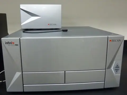
The Infinite M1000 PRO Quadruple Monochromator Microplate Reader (Tecan) is an advanced microplate reader designed for a range of applications, including UV, VIS absorption, fluorescence intensity, and fluorescence polarization. It also supports time-resolved fluorescence, fluorescence resonance energy transfer (FRET), and AlphaScreen technology. The reader can quantify luminescence and analyze luminescence spectra. It features temperature control (up to 42°C) and can shake plates both orbitally and linearly. The system accommodates various plate formats, including 6-, 12-, 24-, 48-, 96-, 384-, and 1536-well plates, and provides high spectral resolution at 1 nm.
-
Image
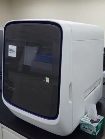
The QuantStudio 12K Flex Real-Time PCR System (Life Technologies) is designed for gene expression analysis, microRNA profiling, and non-coding RNA analysis. It supports both TaqMan and SYBR Green assays and can handle real-time PCR in 96- and 384-well plate formats, as well as TaqMan Low Density Array cards. For more information, click here.
-
Image
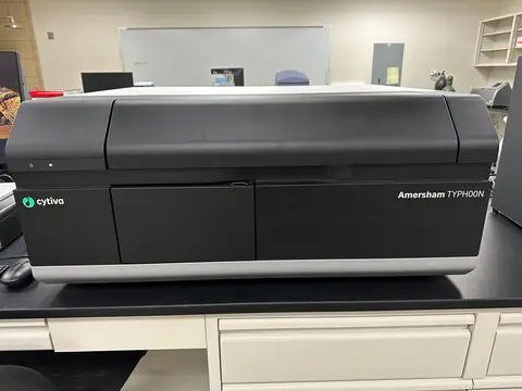
The Typhoon 5 Biomolecular Imager is a versatile imaging system designed for sensitive and quantitative analysis of gels and blots. It excels in phosphoimaging and near-infrared imaging, with capabilities for 700 and 800 nm wavelengths, making it suitable for Western blots with appropriate antibodies. The system features advanced optics for detecting a wide range of fluorescent and chemiluminescent signals. It supports multiple imaging modes, including fluorescent, chemiluminescent, and colorimetric, with customizable resolution settings. For more details, please visit the manufacturer's website.
TTUHSC School of Medicine Cancer Center
The School of Medicine Cancer Center offers multiple core laboratories including the West Texas Pharmacology Core.
The West Texas Pharmacology core facility provides the most up-to-date technologies and expertise in clinical pharmacology in drug development and clinical trials. The core is funded by CPRIT to enable preclinical and clinical pharmacology studies in pediatric cancer drug development and offers the use of a Sciex Triple Quad 6500, Waters Arc HPLC systems (with DAD and FLD), and Shimadzu L20 HPLC.
- Cell Culture Core: Childhood Cancer Repository
- Cancer Animal Models Core
- Cell Line Identification
- Flow Cytometry Core
- Molecular Biology Core
- Pharmacology Core
- Biobanking Core
- Regulatory Sciences Core
- Biostatistics Bioinformatics Core
Texas Tech University
The center provides instrumentation, support and training in various aspects of modern biotechnology including spectrophotometry, protein identification, protein purification molecular interaction, DNA applications and functional genomics.
CASM supports microscopy and cell cytometry research through the use of techniques of transmission electron, scanning electron, laser confocal or epifluorescence microscopy. Trained users are given independent access to the instruments they require.
TTNI is a multi-user neuroimaging facility that promotes cutting-edge interdisciplinary research. It provides researchers with brain and body imaging technologies including structural (MRI), functional magnetic resonance imaging (fMRI), magnetic resonance spectroscopy and diffusion tensor imaging and techniques, including multimodal data fusion of EEG, fMRI, and DTI data.