
Imaging Core Equipment Photos
To use this equipment, contact Dr. Grozdanov at imagecore@ttuhsc.edu or phone: +1 (806) 743-4624.
 |
|
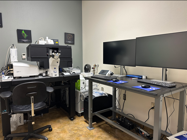
The Zeiss LSM 980 system is a highly advanced confocal microscope designed to enhance both sensitivity and ease of use. Its innovative filterless internal detector layout enables versatile multichannel imaging, complemented by a four-channel GaAsP array that delivers exceptional signal-to-noise ratios for quantitative analysis. The Airyscan 2 detector facilitates rapid and gentle super-resolution imaging based on pixel reassignment. It also facilitates flexible Multiplex modes that allow for the capture of larger fields of view and dynamic biological processes. Key features include: a confocal scanhead with two liquid-cooled MA-PMT detectors and four high-sensitivity GaAsP channels, full emission tunability, and integrated spectral acquisition capabilities. The Zeiss LSM 980 is equipped with solid-state diode laser lines ranging from 405 to 730 nm, including dual-channel near-infrared detectors sensitive to 900 nm. Additional components include an Observer 7 inverted microscope and motorized stand with an automated focus unit, an A.I. Sample Finder, long-lifetime fluorescence LED illumination, and incubation accessories for controlled environments. The system also includes Plan Apochromat objectives, ZEN 3.10 software for advanced imaging capabilities, a PC workstation, and an anti-vibration table, along with a ZEN 3.10 Desk Analysis Software for comprehensive and high-throughput image analysis. |
 |
|
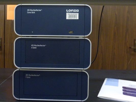
The 4D-Nucleofector Core X/Y (Lonza) system consists of two units: the X Unit, which is designed for transfecting cells in suspension and can handle various cell numbers and formats, and the Y Unit, which is used for transfecting adherent mammalian cells in 24-well plates. Both units are capable of transfecting cells with either DNA or RNA. For more information about the 4D-Nucleofector, please visit the Lonza website. |
 |
|
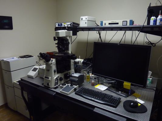
The Nikon T1-E microscope is a versatile imaging system featuring an A1 confocal module and STORM super-resolution capabilities. It can image both live and fixed cells and tissues using conventional or confocal microscopy techniques. Additionally, the STORM module provides super-resolution microscopy, which allows for extremely precise localization of fluorophores beyond the diffraction limit of light, achieving resolution up to ten times greater than standard optical microscopy. The system also supports spectral imaging and advanced fluorescent optical techniques, including TIRF, FRET, and FRAP. It is equipped with a high-resolution EMCCD camera and a stage-top environmental chamber for extended observation of living cells. |
 |
|
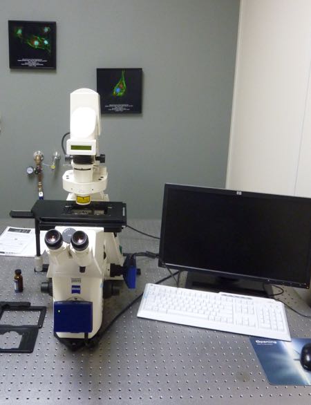
The Zeiss Axiovert 200 M microscope is a popular choice in the Image Analysis Core Facility. This inverted, fully motorized microscope features 10x, 20x, and 63x objectives for wide-field fluorescence microscopy. It has both color and monochrome Axiocam cameras attached, enabling the capture of various samples: color images (e.g., H&E-stained or peroxidase-stained) with the color camera, and low-intensity fluorescent images (e.g., YFP, GFP, RFP, and DAPI) with the monochrome camera. The microscope also supports phase contrast and differential interference contrast (DIC) imaging. Its stage includes a versatile holder that fits different sizes of cell culture dishes and plates, making it easy to quickly assess transfection efficiency. |
 |
|
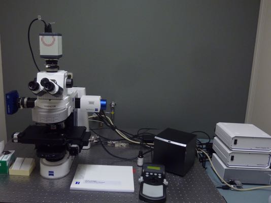
The Zeiss Axio Imager M2 microscope is a fully motorized system designed for brightfield and multi-channel fluorescent imaging. It features motorized x, y, and z stages and is equipped with the Zeiss Apotome system, which uses structured illumination to create sharp optical sections through thick specimens. Images are captured with a low-noise CMOS digital camera (ORCA-Flash4.0, Hamamatsu) with a resolution of 2048 x 2048 pixels. Additionally, the microscope is linked to Zeiss ZEN 3.5 Pro (Blue Edition) software, which offers advanced automated imaging and analysis tools to enhance your research. |
 |
|
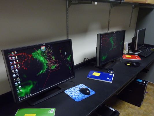
Our workstation area for data analysis is equipped with several computers dedicated to off-line analysis of digital images generated in the facility. Each computer is loaded with essential image analysis software, including Imaris. Imaris offers powerful tools for 3D and 4D image visualization and analysis, allowing you to track and quantify structures within your images. It supports advanced features such as automated object detection, spatial statistics, and detailed visualization of complex datasets. Whether you need to analyze cellular structures, track dynamic processes, or create detailed renderings, Imaris provides a comprehensive suite of tools to enhance your research. |