Imaging Core Lab
Information on Image Core Mission.
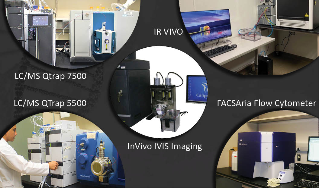
Imaging Core Laboratory Instruments:
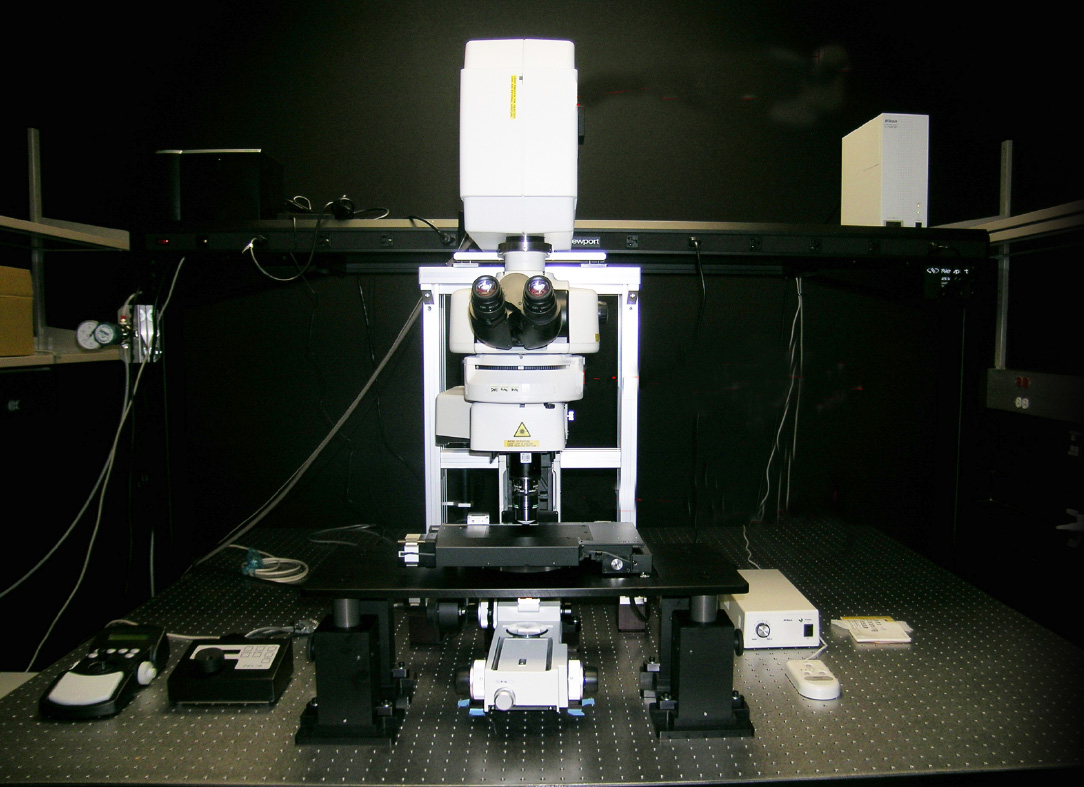
Multiphoton Microscope - Nikon
In live cells and tissue imaging, a key technology is two photon laser scanning microscopy (TPLSM). It minimizes phototoxic effects and offers the possibility to image deep into tissues. A set of instruments using this powerful technology is available in the Imaging Facility at School of Pharmacy.
Key Features:
- Simultaneous excitation imaging with two-wavelength IR laser
- Deep in vivo imaging with super high-sensitive GaAsP NDD
- Ultrahigh-speed imaging of up to 420 frames per second
- Multi-color in vivo imaging
- Deep specimen imaging with non-descanned detectors (NDD)
- Nikon's high-NA objectives are ideal for multiphoton imaging
- Auto laser alignment when changing multiphoton excitation wavelength
- Two types of scanning head enable high-speed, high-quality imaging
- NIS-Elements C acquisition and analysis software
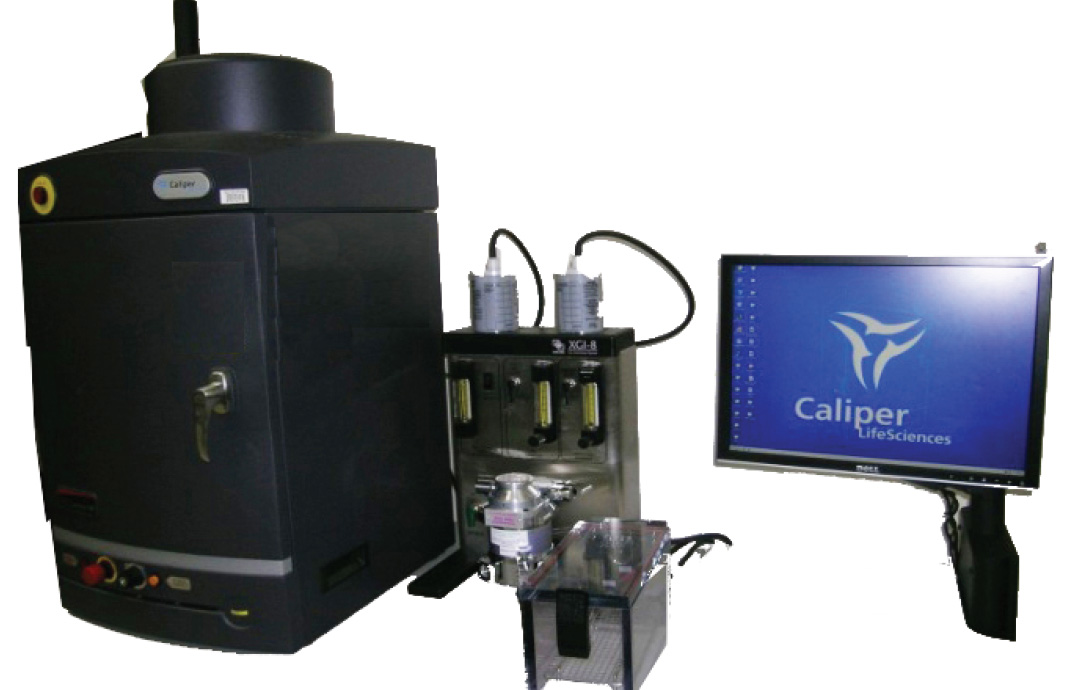
About:
The IVIS Lumina XR system combines highly sensitive bioluminescence and fluorescence
molecular optical imaging with the anatomical precision of digital x-ray. Integrating
these three imaging modalities provides researchers with additional capabilities to
explore the molecular basis of disease and determine the response to drug therapies.
Key Features:
- MULTIMODAL IMAGING :
In vivo preclinical imaging of brain metastases of breast cancer. Left panel shows metastases distribution and location in mouse overlayed on a radiographic image. Scale bar shows areas that have greater tumor burden. Histological verification confirms tumor distribution and location. Imageable metastases were captured approximately four weeks after brain seeking MDA-MB-231 cells were stably transfected with Luc-2 and injected into the circulation of the mouse. (Dr. Paul Lockman) - In Vivo IVIS Lumina XR Imaging
- In Vitro Organ or Tissue Sections
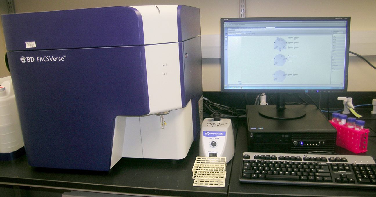
This instrument is equipped with three lasers and can measure up to 10 parameters, offering more options for researchers when designing assays. It is also outfitted with a Flow Sensor, which allows an accurate measurement of cell or particle concentration.
The equipment was purchased with funds provided by Dr. Douglas Stocco and the TTUHSC Research Office, the Departments of Pharmaceutical Sciences and Biomedical Sciences at the School of Pharmacy and the Office of Sciences at the School of Pharmacy. The School of Pharmacy is very excited for this new resource at the Amarillo campus and we wish to thank everyone involved for supporting and advancing this initiative.
Key Features:
Digital Fluorescence-activated cell sorter
2-way sorting and 4-way sorting
Two excitation lasers at 488nm and 633nm wavelength
Updated 06/20/2023
Got Questions?
We're here to help. Contact us if you have questions.
Instrument Usage & Fees
The instruments are available 24/7 except during weekly maintenance or repair periods. Investigators and students must attend a basic training session in order to book and use the equipment.
Online Scheduling:
Individual who have undergone training may book time online through: